- sales@drroyjointclinic.com
- I - 1690, Chittaranjan Park, New Delhi
- Mon - Sat: 10:00 am - 8:00 pm

Gallery Posts


Lorem ipsum dolor sit amet, consectet eiusmod tempor incididunt ut labore e rem ipsum dolor sit amet. sum dolor sit amet, consectet eiusmod.
| Mon - Fri: | 8:00 am - 8:00 pm |
| Saturday: | 9:00 am - 6:00 pm |
| Sunday: | 9:00 am - 6:00 pm |
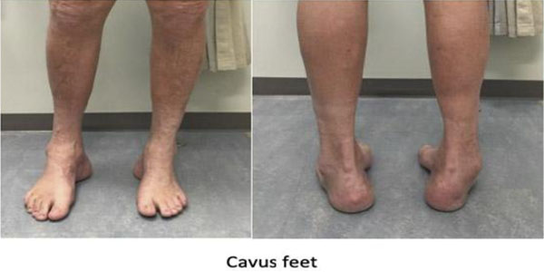
In high arch foot deformity, medial arch is high and most of the sole does not touch the ground and most of the weight bearing happens through heel and ball of the foot. High arch foot is associated with few other foot arch deformity like clawing of toe, heel deformity, plantar fascia contracture, cock-up deformity of great toe.
Most of the time high arch foot deformity is due to some form of neuromuscular deficit either hereditary or acquired both are progressive. It starts developing at puberty and becomes prominent in early adulthood and gradually becomes rigid. Non-neurological one also can be hereditary or acquired and non-progressive.
Neurological:
Non-neurological:
Cavus feet can present with variable features depending on the severity of deformity and causative factor. Pain is a common symptom while walking and standing. Gait is usually unsteady due to inward tilting of heel. Foot drop may happen in the neurological cavus foot due to weakness of the muscles and one can drag his foot while walking. High arch foot is commonly associated with;
Progressive deformity of arch whenever is noticed one should not ignore and contact a specialist Foot & Ankle surgeon. In other stable or subtle condition one should consider visiting a specialist if suffering from
Your doctor will not just examine your feet but also spine, neurological examination, birth history, any cerebrovascular disease and other features for muscular dystrophy. Treatment of high arch foot depends on its causative factor and approach towards neurological and non-neurological cavus feet is totally different. So the main aim of cavus feet diagnosis is directed towards diagnosing the causative neuromuscular disease. He may ask for x-ray of your foot and spine, CT and MRI of your foot. Neurologist evaluation is mandatory for evaluation of neurological aetiology. He may ask for an MRI spine, nerve conduction (NCV), electromyogram (EMG).
Treatment is variable and prognosis also unpredictable depending on severity of deformity and cause of the deformity. Mild deformity with fewer symptoms are managed conservatively and severe deformity requires surgical correction. Mild deformity with associated complications like ankle instability and fracture of 5th metatarsal may require surgical correction of deformity in addition to the treatment of these conditions.
Conservative:
Your doctor will examine your foot to evaluate the severity of deformity as well as its correctability. He will also check the power of different foot muscles as well as range of motion. Will check the other joints for generalised laxity. May enquire about the presence of similar deformity in other siblings or ancestors. He may ask for a weight bearing x-ray of your foot, foot scan. CT and MRI may be required if rigid type flat foot is suspected. USG or MRI is sought for investigation in the early stage of adult acquired flat foot which is called tibialis posterior tendon dysfunction. Although weight bearing x-ray of both feet in both planes is mandatory in every case. Additional heel alignment view is also being done sometimes.
Surgical: Aim of surgical treatment is to get a plantigrade foot to achieve a well distributed weight bearing sole. Surgical procedure is contraindicated in vascular compromise foot. Prognosis is very poor foot with sensory deficit.